Jul 08, 2026
MACSima Imaging System available within core project Z1
Mar 03, 2023
With support of a DFG large equipment grant application, the MACSima ultra high content imaging system was acquired by the WWU in Münster. MACSima is a fully automated fluorescence microscope permitting the analysis of large numbers of epitopes by use of cyclical staining of directly conjugated antibodies. The machine was installed in the middle of February and will be a central part of the Z1 service platform to visualize neutrophils in various tissue environments.
Image
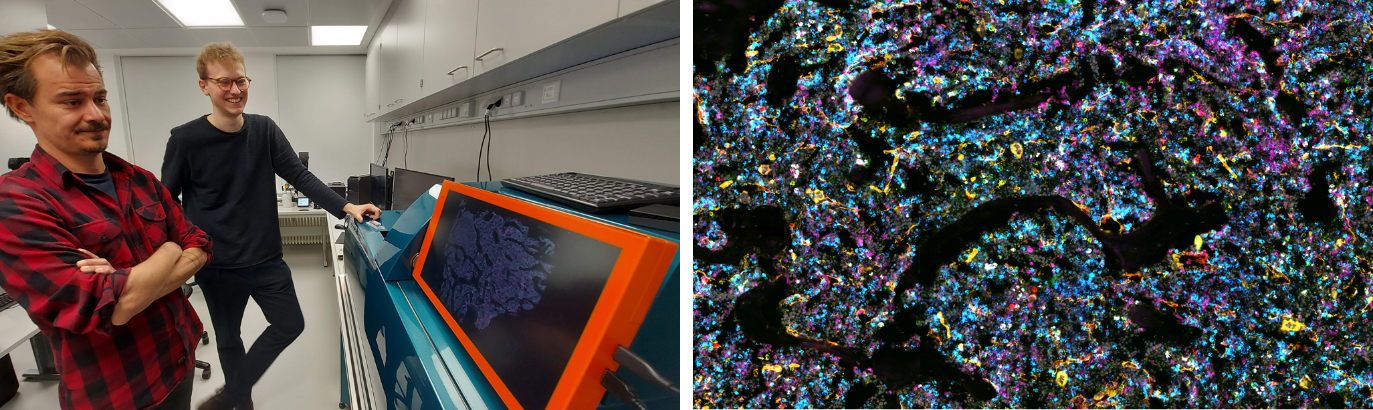
MACSima Imaging system installed at the Institute for Experimental Pathology (ExPat) at the WWU Münster (left). Example of multiplex imaging of the bone marrow (right).